DISCUSSION
Bugsy's owners aren't going to be happy when they find out about the diagnosis: mast cell tumors are generally bad news, though with modern treatments many dogs do survive them, especially if they're spotted in time for effective intervention.
 Mast cells start their lives in the bone marrow, leaving it as monocytes released into the bloodstream. But the monocyte is really just a transit stage: fairly rapidly a monocyte will find a place to exit the vascular system, because they're really a CT cell. They enter intercellular spaces, where they settle down and complete their final differentiation into the definitive mast cell morphology.
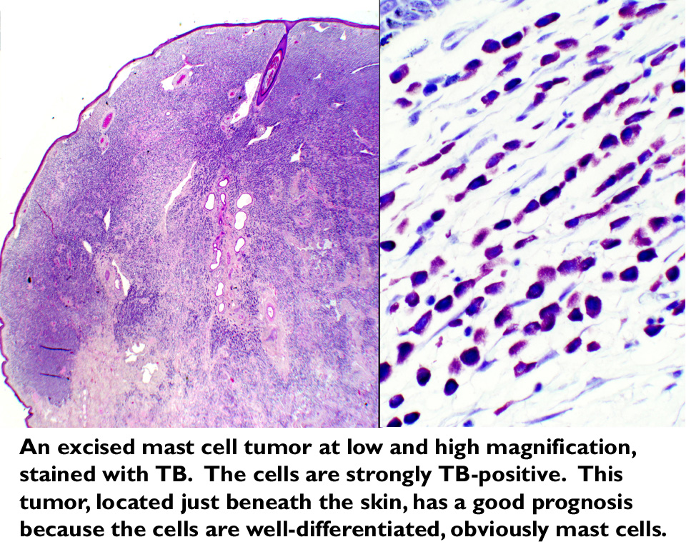
The diagnosis made on the basis of the aspiration is pretty definitive, especially since the vet ordered a toluidine blue stain. TB is specific for mast cell granules.
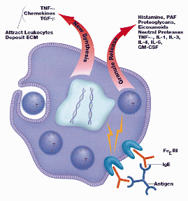
Mast cells are involved in immune reactions to parasitic infection, so Bugsy's owner's guess about an "allergic" reaction isn't too wild, really. When IgE binds to the  mast cell surface, heparin and histamine are released from the granules ("degranulation") into the surrounding tissue. These mediators of inflammation, and other released substances that attract inflammatory cell types, account for the appearance of the site.
The tissue in the vicinity of the bump is inflamed because the cells have inappropriately released their contents. A tumor can have so many mast cells releasing so much material that systemic reactions can occur: vomiting, diarrhea, weight loss, gastric ulceration (manifested as black stools), and in extreme cases, anaphylactic shock. This isn't too likely with a small cutaneous tumor, but MCT's occurring in the deep regions of the body can reach very large size very quickly.
With deep-tissue tumors, sometimes the presenting complaint isn't a "bump" but unexplained malaise. Bugsy's lucky: his tumor is in the dermis (the CT layer of the skin), as most mast cell tumors in dogs tend to be. But a mast cell tumor can develop anywhere there's CT. Mast cells are ubiquitous in their distribution, and you'll find them in any CT including the peritoneal covering of internal organs. MCT's tend to be invasive, shedding tumor cells into lymph nodes and the blood. Cutaneous ones are easier to spot, obviously, and typically less agressive than internal MCTs. Because he gets so much attention, this one was spotted early.
Ultrasound is a quick way to determine if any metastatic tumors have formed yet. Bugsy is lucky: his tumor is small and hasn't seeded new ones. Nor does he have circulating mast cells in his blood (a condition of mastocytosis, a very bad sign).
Nobody knows what causes MCTs. What triggers a single mast cell to begin the process of dividing and creating a clone? Certainly, there are genetic factors involved: breed dispositions to MCTs are well known (English Bulldogs are one of the breeds at risk, but any dog can develop MCTs) but undoubtedly there are other triggers: exposure to carcinogens, localized increases in mast cell populations due to irritations, and so forth. Nobody really has an answer—yet.
Mast cell tumors should be removed immediately. Unfortunately, getting all of the tumor isn't always possible. Tumors that have reached a significant size form octopus-like arms, and getting clean margins requires a pretty wide excision. Bugsy's in for a couple of weeks wearing a cone on his head. Surgical removal is the first line of treatment: many vets follow this up with radiation therapy to destroy any missed cells; in cases where spread has happened, chemotherapy with corticosteroids and cytotoxic drugs has to be considered.
References:
Bonagura, J. Current Veterinary Therapy 12. W.B. Saunders Co. Philadelphia, PA; 1995.
Chun, R. Canine cutaneous mast cell tumors: One of these is not like the other. Presented at the Western Veterinary Conference, 2004. Las Vegas, Nevada.
Rogers, KS. Mast Cell Disease. In Ettinger, SJ; Feldman EC (eds.) Textbook of Veterinary Medicine. W.B. Saunders Co. Philadelphia, PA; 2005: 773-778.
Piscopo, S. Canine Mast Cell Tumors. Veterinary Forum. June, 1999.
Vail, D. Dealing with Canine Mast Cell Tumors. Veterinary Product News. December, 1999.
 Mast cells start their lives in the bone marrow, leaving it as monocytes released into the bloodstream. But the monocyte is really just a transit stage: fairly rapidly a monocyte will find a place to exit the vascular system, because they're really a CT cell. They enter intercellular spaces, where they settle down and complete their final differentiation into the definitive mast cell morphology.
  mast cell surface,